Iliotibial Band Syndrome (ITBS)

October 7, 2025

Causes, Symptoms & Treatment | Weaver Physiotherapy Northwich

Understanding Iliotibial Band Syndrome


If you’re a runner, cyclist, or active individual experiencing pain on the outer side of your knee or thigh, you could be dealing with Iliotibial Band Syndrome (ITBS) — one of the most common causes of lateral knee pain in athletes.


At Weaver Physiotherapy & Sports Injury Clinic in Northwich, Cheshire, our team of Chartered Physiotherapists specialises in diagnosing and treating Iliotibial Band Syndrome, helping runners and athletes of all levels recover quickly, move freely, and prevent recurrence.


With over 70 years of combined clinical experience, our physiotherapists combine hands-on treatment, advanced rehabilitation, and movement re-education to tackle ITBS at its source.


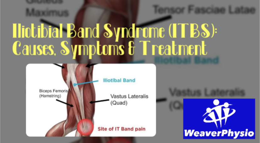
What Is the Iliotibial Band?


The iliotibial band (ITB) is a thick band of connective tissue that runs from the hip to the outer side of the knee, connecting the tensor fasciae latae (TFL) and gluteus maximus muscles to the tibia (shin bone). Its role is to stabilise the knee and hip during movement, particularly when running, walking, or cycling.


When functioning properly, the IT band glides smoothly over the bony structures around the knee. However, if it becomes tight, inflamed, or irritated from repetitive friction, it can cause pain on the outside of the knee — the hallmark of Iliotibial Band Syndrome.


What Causes Iliotibial Band Syndrome?


ITBS is an overuse injury, meaning it develops gradually from repetitive stress rather than a single traumatic event. The condition is especially common among runners, cyclists, hikers, and gym-goers who perform frequent bending and straightening of the knee.


Common Causes Include:

    1.    Overtraining or Sudden Increase in Activity

Increasing your mileage, pace, or hill training too quickly can overload the IT band and surrounding structures.

    2.    Muscle Imbalances and Weakness

Weak gluteal or hip stabilising muscles cause the IT band to take on excessive tension, leading to irritation around the knee.

    3.    Tightness in the IT Band or Hip Muscles

Limited flexibility in the TFL, glutes, or quadriceps can increase friction along the IT band.

    4.    Poor Running Form or Biomechanics

Excessive inward knee movement (valgus), overstriding, or poor hip control can increase stress on the outer knee.

    5.    Improper Footwear or Running Surface

Worn-out shoes, excessive camber (running on sloped surfaces), or running always in one direction can contribute to ITBS.

    6.    Leg Length Discrepancy or Pelvic Alignment Issues

Subtle biomechanical differences can lead to uneven stress distribution across the knees and hips.


Who Is Most at Risk?


Iliotibial Band Syndrome can affect anyone who is active, but some groups are particularly prone to it:

    •    Distance runners, especially those training for marathons or half-marathons

    •    Cyclists with poor bike setup or excessive mileage

    •    Hikers tackling long or uneven terrain

    •    Footballers, rugby players, and gym-goers doing repetitive squatting or lunging

    •    Office workers with tight hip flexors or glutes due to prolonged sitting


If you’re training hard but developing sharp or aching pain on the outside of your knee, you may be in the early stages of ITBS.


Symptoms of Iliotibial Band Syndrome


The hallmark symptom of ITBS is pain on the outer (lateral) side of the knee, but the condition can cause a range of sensations and discomfort patterns.


Common Symptoms Include:

    •    Sharp or burning pain on the outside of the knee

    •    Pain that worsens with running, especially downhill or after several miles

    •    Discomfort when climbing stairs or getting up from a seated position

    •    Tightness or tenderness along the outer thigh

    •    Snapping or clicking sensation around the knee

    •    Pain that may radiate up towards the hip or down the leg


The pain often starts gradually and worsens with continued activity. Early intervention is crucial to prevent the condition from becoming chronic.


How Is Iliotibial Band Syndrome Diagnosed?


At Weaver Physiotherapy, our expert clinicians take a comprehensive approach to diagnosis. We don’t just treat the pain — we identify the underlying cause.


Our ITBS Assessment Includes:

    1.    Detailed Case History

Understanding your training habits, footwear, injury history, and lifestyle factors.

    2.    Physical Examination

Assessing hip, knee, and ankle alignment, as well as muscle strength and flexibility.

    3.    Movement Analysis

Evaluating running gait, walking pattern, or cycling posture to identify mechanical contributors.

    4.    Palpation & Orthopaedic Tests

Testing specific structures to rule out other causes of knee pain (e.g., meniscus injury, lateral ligament strain).

    5.    Video Gait Analysis

Using slow-motion technology to spot subtle biomechanical issues that lead to ITB irritation.


This holistic assessment ensures your treatment plan is tailored to your individual needs and the root cause of your problem.


Physiotherapy Treatment for Iliotibial Band Syndrome


1. Pain Relief and Inflammation Management


The first goal is to reduce pain and inflammation. Your physiotherapist may use:

    •    Manual therapy and soft tissue release to ease tension along the IT band and surrounding muscles.

    •    Ice therapy or ultrasound for inflammation control.

    •    Kinesiology taping to offload the IT band and support healing.


2. Deep Tissue and Sports Massage


Massage therapy is extremely effective in reducing IT band tightness. At Weaver Physio, our Sports Massage and Deep Tissue Massage treatments target the TFL, glutes, quadriceps, and lateral thigh to restore normal tissue mobility and reduce friction at the knee.


3. Shockwave Therapy


For chronic or stubborn cases, Shockwave Therapy provides a powerful, evidence-based solution. It uses acoustic waves to stimulate blood flow, break down scar tissue, and accelerate healing in the IT band and surrounding tendons. NICE (National Institute for Health and Care Excellence) recommends this treatment for persistent soft tissue conditions.


4. Strength & Conditioning Rehabilitation


Once pain is under control, strengthening exercises are essential to correct underlying imbalances. Your physiotherapist will design a tailored exercise program targeting:

    •    Gluteus medius and maximus (hip stabilisers)

    •    Core muscles (for pelvic control)

    •    Hamstrings and quadriceps (to balance knee mechanics)


5. Gait Re-Education


Poor running form often drives ITBS. Our Runner’s MOT and Video Gait Analysis sessions identify biomechanical inefficiencies, helping you adjust stride length, cadence, and posture for long-term prevention.


6. Stretching and Mobility Work


Specific stretches for the IT band, TFL, glutes, and quads help relieve tension and maintain flexibility. Foam rolling and mobility drills may also be prescribed.


7. Return to Sport Planning


We guide you safely back into training with progressive loading, ensuring your tissue capacity matches your running or cycling demands.


At Weaver Physio, your treatment plan is completely individualised — addressing pain relief, restoring function, and preventing recurrence.


Exercises for Iliotibial Band Syndrome


Here are a few examples of physiotherapy-approved exercises often included in recovery plans:

    1.    Side-Lying Leg Raises – Strengthens the gluteus medius.

    2.    Clamshells – Targets hip stabilisers and improves knee alignment.

    3.    Hip Bridges – Builds glute strength and reduces ITB tension.

    4.    Foam Rolling the IT Band – Helps release tight fascia and improve mobility.

    5.    Standing IT Band Stretch – Gently lengthens the lateral thigh structures.


(Always consult your physiotherapist before starting new exercises, as incorrect technique can worsen symptoms.)


How Long Does Recovery Take?


Recovery time depends on severity, chronicity, and training load, but most patients improve significantly within 4–8 weeks of structured physiotherapy and activity modification. Chronic or long-standing cases may take longer, especially if biomechanical retraining is needed.


Our goal at Weaver Physio is to not only get you pain-free, but also to build resilience so you can return to running, cycling, or sport with confidence — and without fear of re-injury.


Preventing Iliotibial Band Syndrome


1. Gradual Training Progression


Avoid sudden increases in distance, pace, or hill work. Follow the 10% rule – increase weekly mileage by no more than 10%.


2. Strengthen Your Glutes & Core


Regular hip and core strengthening reduces ITB strain during movement.


3. Prioritise Flexibility


Incorporate regular stretching and foam rolling of the IT band, quadriceps, hamstrings, and calves.


4. Check Your Footwear


Replace worn-out shoes and ensure your trainers match your running style. Gait analysis can identify whether you need neutral or stability shoes.


5. Cross-Train & Rest


Include low-impact activities like swimming or cycling, and schedule adequate rest days to allow tissue recovery.


6. Get a Professional Running Assessment


Our Runner’s MOT identifies inefficiencies in your stride and recommends personalised corrective exercises — ideal for injury prevention and performance enhancement.


Why Choose Weaver Physiotherapy for IT Band Syndrome Treatment?


At Weaver Physiotherapy & Sports Injury Clinic, we’re passionate about helping runners, athletes, and active individuals overcome injury and return to what they love.


What Sets Us Apart:


✅ Specialist Running Injury Clinic – Experts in ITBS, runner’s knee, Achilles pain, and shin splints

✅ 70+ Years Combined Experience – Trusted Chartered Physiotherapists in Cheshire

✅ Comprehensive Assessment – Including video gait analysis and functional testing

✅ Advanced Treatments – Shockwave therapy, acupuncture, sports massage, rehab & manual therapy

✅ Tailored Rehabilitation Plans – Designed around your sport, goals, and lifestyle

✅ Local, Trusted Experts – Serving Northwich, Knutsford, Winsford, Middlewich, Frodsham & Tarporley


Whether you’re a Parkrunner, marathoner, or simply love staying active, our mission is to help you move better, perform better, and live pain-free.


Take Control of Your Recovery Today


Don’t let IT band pain stop you from doing what you love. Early intervention is key — the sooner you seek help, the faster you’ll recover.


📞 Call Weaver Physiotherapy & Sports Injury Clinic today on 01606 227484

🌐 Book online at https://www.weaverphysio.com

📍 Weaver Physio – Your Trusted Partner in Recovery, Performance & Pain-Free Living


October 7, 2025
Expert Advice from Weaver Physiotherapy, Northwich
October 7, 2025
Causes, Symptoms & Effective Treatment | Weaver Physiotherapy, Northwich
October 6, 2025
Causes, Symptoms & Physiotherapy Treatment in Cheshire
October 6, 2025
Expert Advice from Weaver Physiotherapy & Sports Injury Clinic, Northwich
October 5, 2025
Knee pain is one of the most common musculoskeletal complaints we see at Weaver Physiotherapy & Sports Injury Clinic in Northwich, Cheshire. From runners and athletes to office workers and retirees, knee problems can affect anyone. While many people are familiar with conditions such as arthritis, meniscus tears, or ligament injuries, one often-overlooked cause of knee swelling and discomfort is a Baker’s Cyst. Also known as a popliteal cyst, this condition can cause swelling behind the knee, stiffness, and reduced mobility – and if untreated, it may lead to ongoing pain or limit your ability to exercise, walk comfortably, or perform daily activities. In this in-depth guide, we’ll explain what a Baker’s Cyst is, the symptoms to look out for, why it develops, and most importantly – how physiotherapy can help you manage, treat, and prevent it. What is a Baker’s Cyst? A Baker’s Cyst is a fluid-filled swelling that develops behind the knee, in the popliteal space. It occurs when excess joint fluid (synovial fluid) escapes from the knee joint and collects in a small sac behind the knee. This fluid build-up is often a result of an underlying knee problem, such as: • Osteoarthritis (wear and tear of the knee joint) • Rheumatoid arthritis • Meniscus injuries (cartilage tears) • Ligament injuries • Knee joint inflammation The cyst itself is not dangerous, but it can be painful, restrict movement, and cause the back of the knee to feel tight – especially when bending or straightening the leg. Symptoms of a Baker’s Cyst Some people with a Baker’s Cyst may not experience symptoms at all, especially if the cyst is small. However, when symptoms do occur, they may include: ✔️ A noticeable lump or swelling behind the knee ✔️ Tightness or stiffness in the back of the knee ✔️ Pain or aching, particularly when straightening or bending the leg ✔️ Reduced flexibility and mobility ✔️ Swelling in the calf (if the cyst bursts and fluid leaks down the leg) In rare cases, a ruptured Baker’s Cyst can mimic the symptoms of a blood clot (deep vein thrombosis – DVT), with sudden calf swelling, pain, and redness. If you experience these symptoms, urgent medical assessment is required. Causes: Why Do Baker’s Cysts Develop? A Baker’s Cyst does not develop in isolation – it usually results from knee joint changes or injury that causes excess fluid production. Common causes include: 1. Arthritis • Osteoarthritis is a major cause. As the cartilage wears down, the joint becomes inflamed, leading to fluid build-up. • Rheumatoid arthritis, an autoimmune condition, can also trigger joint swelling and cyst formation. 2. Sports Injuries • Damage to the meniscus (cartilage) can irritate the joint and lead to cysts. • Ligament injuries (ACL, MCL) may also increase fluid build-up. 3. Overuse or Repetitive Strain • Runners, cyclists, and people who frequently squat or kneel may place repetitive stress on the knee joint, contributing to cyst formation. 4. Underlying Inflammation • Any condition that causes inflammation within the knee joint (including gout) can lead to synovial fluid leakage and cyst development. Diagnosis of a Baker’s Cyst At Weaver Physiotherapy & Sports Injury Clinic, our Chartered Physiotherapists carry out a detailed assessment to identify whether your knee pain and swelling are caused by a Baker’s Cyst, or another condition. The process may include: 🔹 Clinical assessment – checking your symptoms, medical history, and examining the back of the knee for swelling. 🔹 Ultrasound scan – often recommended to confirm the diagnosis. 🔹 MRI scan – if an underlying injury such as a cartilage tear is suspected. Early diagnosis is key. Many patients believe they just have “knee swelling” without realising it’s linked to an underlying joint issue. How Physiotherapy Helps Baker’s Cyst Physiotherapy is one of the most effective ways to manage and treat a Baker’s Cyst. While draining the cyst or surgery is sometimes required in severe cases, the majority of patients benefit from conservative treatment that targets the root cause. At Weaver Physio, our approach focuses on: 1. Pain Relief & Swelling Reduction • Manual therapy to ease stiffness • Ice, compression, and elevation strategies • Advice on activity modification to avoid aggravation 2. Improving Knee Mobility • Gentle stretching exercises to restore range of movement • Targeted flexibility work for the hamstrings and calf muscles 3. Strengthening Exercises • Quadriceps strengthening to improve joint support • Glute activation to reduce stress on the knee • Core stability to enhance overall lower-limb mechanics 4. Correcting Biomechanics • Video Gait Analysis and biomechanical assessment (part of our Runner’s MOT service) • Identifying imbalances in walking or running that may contribute to knee overload 5. Treating the Underlying Cause • If arthritis is the cause, we’ll develop a long-term joint management plan • If it’s due to a sports injury, we’ll provide a tailored rehabilitation programme Home Management Strategies Alongside professional physiotherapy, patients can often manage symptoms at home with: ✔️ Rest & Activity Modification – avoiding deep squats or repetitive knee strain ✔️ Ice Therapy – applying ice packs to reduce swelling ✔️ Compression Supports – knee braces can help control fluid build-up ✔️ Gentle Exercises – as advised by your physiotherapist When is Surgery Needed? Surgery is rarely the first option for Baker’s Cyst. It is usually only considered if: • The cyst is very large and painful • Conservative treatments have failed • The underlying cause (such as a meniscus tear) requires surgical repair In such cases, a surgeon may drain the cyst (aspiration) or repair the damaged structures inside the knee. Can a Baker’s Cyst Burst? Yes. In some cases, the cyst can rupture, releasing fluid into the calf. This can cause sudden pain, swelling, and bruising in the lower leg. It may resemble a DVT (blood clot), so it’s important to seek urgent medical advice to rule this out. Recovery Timeline Recovery depends on the underlying cause: • Minor cases: Symptoms may improve in a few weeks with rest and physiotherapy. • Arthritis-related cases: Long-term management is required, but physiotherapy helps control pain and mobility. • Sports injuries: Recovery may take 6–12 weeks, depending on the severity of the meniscus or ligament damage. At Weaver Physio, we’ll give you a personalised recovery plan so you know exactly what to expect at each stage. Preventing Baker’s Cyst While not all cases can be prevented, you can reduce your risk by: 🔹 Maintaining strong quadriceps, hamstrings, and glutes 🔹 Avoiding repetitive high-impact activities without proper recovery 🔹 Wearing appropriate footwear for your activity 🔹 Seeking physiotherapy early if you experience knee swelling or stiffness Why Choose Weaver Physio for Knee Pain? At Weaver Physiotherapy & Sports Injury Clinic in Northwich, Cheshire, we are specialists in diagnosing and treating knee injuries and conditions such as Baker’s Cyst. Here’s why patients across Cheshire – including Northwich, Knutsford, Winsford, Middlewich, Tarporley, and Frodsham – choose us: ✔️ 70+ years of combined clinical experience ✔️ Experts in sports injuries, arthritis, and musculoskeletal pain ✔️ Access to advanced services such as Shockwave Therapy, Acupuncture & Video Gait Analysis ✔️ Tailored rehabilitation plans that get to the root cause of your pain ✔️ Trusted by runners, athletes, and the local community Our goal is simple: to help you recover faster, prevent future injuries, and get back to living pain-free. Book Your Appointment If you’re struggling with knee pain or suspect you may have a Baker’s Cyst, don’t ignore the symptoms. Early treatment can make a huge difference in your recovery and long-term knee health. 📞 Call us today on 01606 227484 🌐 Visit us at http://www.weaverphysio.com Weaver Physiotherapy & Sports Injury Clinic – Your Trusted Partner in Recovery, Performance & Pain-Free Living.
October 3, 2025
Causes, Symptoms & Physiotherapy Treatment
October 3, 2025
Causes, Symptoms, Diagnosis & Treatment | Weaver Physiotherapy & Sports Injury Clinic, Cheshire
October 2, 2025
Causes, Symptoms & Physiotherapy Treatment | Weaver Physiotherapy Northwich
October 1, 2025
Shoulder pain is one of the most common musculoskeletal complaints we see at Weaver Physiotherapy & Sports Injury Clinic in Northwich, Cheshire. Among the many conditions that can affect the shoulder, Long Head of Biceps Tendinitis (LHBT) is a frequent source of pain, stiffness, and reduced function – especially in athletes and active individuals. If you’ve noticed pain at the front of your shoulder that worsens with lifting, reaching, or overhead activity, you may be experiencing inflammation of the biceps tendon. The good news is, with early diagnosis and expert physiotherapy management, this condition can be successfully treated – allowing you to get back to sport, training, and everyday life without pain. In this blog, we’ll explain: • What the long head of the biceps tendon is • The main causes and risk factors for tendinitis • Common symptoms and warning signs • How it is diagnosed • Treatment and rehabilitation options at Weaver Physio • Tips to prevent recurrence and protect your shoulder Understanding the Long Head of the Biceps Tendon The biceps muscle has two tendons at the shoulder: • Short head – attaches to the coracoid process at the front of the shoulder blade. • Long head – runs through the shoulder joint itself, attaching to the top of the glenoid (shoulder socket). Because the long head of the biceps tendon passes through a narrow groove and is closely associated with the rotator cuff, it is more vulnerable to irritation, inflammation, and injury. Its function is to: • Assist in shoulder flexion (lifting the arm forward). • Contribute to shoulder stability, particularly with overhead movements. • Aid in supination (turning the palm upwards). When overloaded or irritated, the tendon can become inflamed, leading to tendinitis. If not treated properly, this may progress to tendon degeneration (tendinopathy) or even partial tears. Causes of Long Head of Biceps Tendinitis At Weaver Physio, we often see long head of biceps tendinitis linked to repetitive overuse, poor biomechanics, or secondary shoulder problems. 1. Overuse & Repetitive Movements • Repeated overhead activity (e.g. swimming, tennis, cricket, weightlifting). • Heavy lifting in the gym without correct technique. • Throwing sports that stress the tendon. 2. Shoulder Impingement & Rotator Cuff Dysfunction The biceps tendon often works in partnership with the rotator cuff. Weakness, imbalance, or injury in the cuff can increase strain on the tendon. 3. Poor Posture Rounded shoulders and forward head posture (common in office workers) can narrow the bicipital groove, irritating the tendon. 4. Age-Related Degeneration As we age, tendons naturally lose some elasticity and become more prone to inflammation or microtears. 5. Direct Trauma A fall, awkward lifting, or sudden jerk to the arm may trigger tendinitis. Symptoms of Long Head of Biceps Tendinitis The hallmark symptom is pain at the front of the shoulder, but several features can help identify LHBT tendinitis: • Sharp or aching pain at the anterior shoulder. • Pain aggravated by lifting, carrying, or overhead reaching. • Tenderness when pressing on the bicipital groove (front of the shoulder). • Discomfort when turning the palm upward against resistance (supination). • Possible clicking or catching sensation in the shoulder. • Weakness when lifting objects or performing overhead movements. In severe or chronic cases, swelling and reduced range of motion may also be present. Diagnosing Biceps Tendinitis At Weaver Physiotherapy & Sports Injury Clinic, our expert clinicians use a combination of: • Clinical history & symptom analysis – understanding your activity, pain pattern, and injury history. • Physical examination – testing shoulder movement, strength, and palpating the tendon. • Special tests – such as Speed’s test or Yergason’s test to reproduce symptoms. • Referral for imaging (if required) – ultrasound or MRI may be suggested to rule out rotator cuff tears, labral pathology, or joint issues. Accurate diagnosis is essential, as anterior shoulder pain may also be caused by conditions like rotator cuff tendinopathy, impingement syndrome, labral tears, or osteoarthritis. Treatment for Long Head of Biceps Tendinitis At Weaver Physio, we provide individualised, evidence-based treatment plans designed to: 1. Reduce pain and inflammation. 2. Restore shoulder movement and strength. 3. Correct underlying biomechanical issues. 4. Prevent recurrence and future injury. 1. Early Stage: Pain Relief & Inflammation Control • Rest & activity modification – avoiding aggravating movements. • Ice therapy – reduces acute inflammation. • Manual therapy – soft tissue release, joint mobilisation. • Taping techniques – to offload the tendon. 2. Physiotherapy & Rehabilitation • Targeted strengthening – rotator cuff, scapular stabilisers, and core muscles. • Stretching & mobility exercises – to restore range and reduce stiffness. • Postural correction – improving alignment to reduce tendon stress. • Progressive loading – gradually strengthening the tendon to handle daily and sporting demands. 3. Adjunct Treatments • Shockwave Therapy – highly effective for chronic or stubborn tendon pain, stimulating healing. • Acupuncture / Dry Needling – reduces pain and improves circulation. • Sports & Deep Tissue Massage – eases tightness and improves blood flow. 4. Return to Sport & Prevention • Video Gait & Movement Analysis – identifies biomechanical flaws. • Strength & Conditioning Programmes – tailored to sport demands. • Education & Training Advice – ensuring safe progression back to activity. Recovery Timeline Recovery depends on severity and treatment compliance, but typical timelines are: • Mild tendinitis – 3 to 6 weeks with physiotherapy. • Moderate cases – 6 to 12 weeks with progressive loading. • Chronic / degenerative tendinopathy – 3 to 6 months, sometimes longer if associated with rotator cuff tears. At Weaver Physio, our aim is not just pain relief but long-term recovery, ensuring you return to sport stronger and more resilient. Preventing Biceps Tendinitis Prevention is always better than cure. Our physiotherapists recommend: • Warm up properly before sport or gym sessions. • Strengthen rotator cuff and scapular stabilisers. • Avoid excessive repetitive overhead movements. • Correct poor posture, especially if working at a desk. • Progress training gradually – avoid sudden load spikes. • Include mobility and stretching in your routine. Why Choose Weaver Physio for Shoulder & Sports Injuries? At Weaver Physiotherapy & Sports Injury Clinic in Northwich, we pride ourselves on being Cheshire’s Sports Injury Specialists. • 🏆 70+ years of combined clinical experience • 🩺 Chartered Physiotherapists with advanced expertise in sports injuries • 💪 Evidence-based treatment plans tailored to your body and goals • 🌀 Specialist services including Shockwave Therapy, Sports Massage, Acupuncture, and Gait Analysis • 📍 Conveniently located in Northwich, supporting patients from Knutsford, Winsford, Middlewich, Frodsham, Tarporley, and across Cheshire We help athletes, gym-goers, office workers, and active individuals recover faster, prevent setbacks, and stay pain-free. Book Your Appointment Today If shoulder pain is holding you back, don’t wait for it to get worse. Early treatment is the key to faster recovery and avoiding long-term damage. 👉 Call 01606 227484 to book your appointment. 👉 Visit www.weaverphysio.com to learn more. At Weaver Physio, we are your trusted partner in recovery, performance, and pain-free living.
October 1, 2025
At Weaver Physiotherapy & Sports Injury Clinic in Northwich, Cheshire, our Chartered Physiotherapists treat a wide range of wrist and hand injuries. One of the lesser-known but increasingly common conditions is a TFCC tear – short for Triangular Fibrocartilage Complex tear. This type of injury often causes persistent wrist pain, weakness, and reduced grip strength, and if not managed properly, it can affect everyday activities, work, and sport. In this comprehensive guide, we’ll explore: • What the TFCC is and why it’s important • Common causes and risk factors for TFCC tears • Key symptoms and warning signs • How TFCC tears are diagnosed • Effective treatment options including physiotherapy • How Weaver Physio can help you recover and get back to doing what you love What is the TFCC? The Triangular Fibrocartilage Complex (TFCC) is a structure located on the small-finger (ulnar) side of the wrist. It’s made up of cartilage and ligaments that: • Stabilise the wrist joint • Cushion and absorb shock between the forearm bones (ulna and radius) • Support gripping, lifting, twisting, and weight-bearing through the wrist Because of its role in stabilising and distributing force, the TFCC is vulnerable to injury – especially during sports, repetitive hand movements, or a fall on an outstretched hand. Causes of a TFCC Tear There are two main types of TFCC tears: traumatic and degenerative. 1. Traumatic TFCC Tear These are caused by sudden injury or impact, often involving excessive twisting or loading of the wrist. Common examples include: • Falling on an outstretched hand (FOOSH injury) • Sports injuries in activities like tennis, gymnastics, football, or rugby • Heavy lifting or sudden pulling actions • Car accidents or direct impact to the wrist 2. Degenerative TFCC Tear These tears develop gradually over time due to natural wear and tear or repetitive stress. Risk factors include: • Repetitive wrist use (e.g. manual labour, factory work, trades) • Age-related degeneration of cartilage • Ulnar variance (when the ulna bone is slightly longer than the radius, increasing TFCC load) • Previous wrist injuries leading to instability Symptoms of a TFCC Tear The symptoms can vary depending on severity, but common signs include: • Ulnar-sided wrist pain (pain on the little finger side of the wrist) • Pain when twisting the wrist (turning a doorknob, using a screwdriver, opening jars) • Clicking, popping, or grinding sensations in the wrist • Reduced grip strength and difficulty lifting objects • Swelling or tenderness around the wrist joint • Pain during weight-bearing activities such as pushing up from a chair or doing push-ups • In severe cases, instability or weakness when gripping or rotating the wrist If left untreated, a TFCC tear can become a long-term problem, affecting daily activities, sport, and work. Diagnosing a TFCC Tear Accurate diagnosis is vital for effective treatment. At Weaver Physio, our expert team combines clinical assessment with referral for imaging if needed. 1. Clinical Examination • Detailed history of how the pain started • Wrist palpation and movement testing • Special tests such as the ulnar grind test or TFCC load test 2. Imaging Tests • MRI scan is the gold standard for detecting TFCC tears • X-rays may be used to rule out fractures or joint abnormalities • Ultrasound can sometimes help assess soft tissue injuries Treatment for TFCC Tears The treatment plan depends on the severity of the tear, the patient’s lifestyle, and whether the tear is traumatic or degenerative. 1. Conservative (Non-Surgical) Management Most TFCC tears respond well to non-surgical treatment – especially when managed early. • Rest & Activity Modification – avoiding activities that aggravate the wrist • Splinting or Bracing – to immobilise and allow healing • Ice & Pain Relief – reducing pain and inflammation • Physiotherapy – targeted exercises, manual therapy, and progressive rehabilitation • Corticosteroid injections – occasionally used for stubborn inflammation 2. Physiotherapy for TFCC Tears at Weaver Physio At Weaver Physiotherapy & Sports Injury Clinic, we specialise in tailored rehabilitation programmes for wrist injuries. Treatment may include: ✔️ Hands-on therapy to improve mobility and reduce stiffness ✔️ Strengthening exercises for forearm, grip, and wrist stability ✔️ Range of motion exercises to restore flexibility ✔️ Proprioception and coordination training for wrist control ✔️ Sport-specific or work-related rehabilitation to ensure safe return 3. Surgical Management If the tear is severe, unstable, or does not respond to conservative care, referral to a specialist may be necessary. Surgery may involve: • Arthroscopic debridement (cleaning up damaged tissue) • Repair of the TFCC using sutures • Ulnar shortening procedures if bone length contributes to tearing Post-surgical recovery still requires structured physiotherapy to regain strength, mobility, and function. Recovery Timeline for TFCC Tears Recovery varies depending on the severity of the injury and whether surgery is required: • Mild tears: 4–6 weeks with physiotherapy and bracing • Moderate tears: 8–12 weeks of structured rehab • Post-surgery: 3–6 months for full recovery and return to sport/work At Weaver Physio, we create an individualised recovery plan to guide you through every stage – from early pain relief to advanced rehabilitation and return to activity. Preventing TFCC Tears While not all injuries can be prevented, you can reduce risk by: • Avoiding repetitive heavy wrist strain where possible • Using proper wrist support in high-impact sports • Strengthening forearm and wrist muscles • Addressing early signs of pain before they worsen Why Choose Weaver Physiotherapy for TFCC Tear Treatment? At Weaver Physiotherapy & Sports Injury Clinic in Northwich, we are trusted by patients across Knutsford, Winsford, Middlewich, Tarporley, Frodsham, and Cheshire for expert assessment and treatment of wrist injuries. Here’s why patients choose us: • 70+ years of combined clinical experience • Chartered Physiotherapists with advanced skills in sports injuries, musculoskeletal rehab, and pain management • Access to advanced treatments including shockwave therapy, acupuncture, and tailored rehabilitation • A patient-centred approach focused on long-term recovery, not just short-term relief • Convenient clinic location, flexible appointments, and personalised care Whether you’re an athlete, manual worker, or simply someone struggling with wrist pain, we’ll help you recover, prevent further problems, and get back to living pain-free. Final Thoughts A TFCC tear can be painful, frustrating, and limiting – but with the right diagnosis and treatment, recovery is absolutely possible. Early intervention, physiotherapy, and guided rehabilitation make a huge difference to outcomes. If you’re struggling with persistent wrist pain, clicking, or weakness, don’t ignore it. The earlier you seek treatment, the faster and more effective your recovery will be. 📞 Call 01606 227484 today or visit 🌐 www.weaverphysio.com to book your consultation at Weaver Physiotherapy & Sports Injury Clinic – Your Trusted Partner in Recovery, Performance & Pain-Free Living.
Show More